
Sophia Ogot
Coronado High School
Class Of 2020
MRI & EEG Experience
Synthesized New Knowledge Of
Neuroscience Into A Proposal
For A Neuroscience Experiment
Spectroscopy To Measure Brain
Conducted A Near-Infrared
Activity Of A Live Person
My Neuroscience Training
The Future
Moving Towards
Completed (Click Here To Learn More)
Training With Leading
Neuroscience Experts
Completed (Click Here To Learn More)
Neuroscience
Experimentation
Completed (Click Here To Learn More)
Completed (Click Here To Learn More)
MRI & EEG
Experience
1
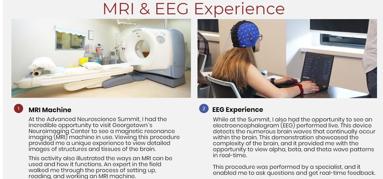
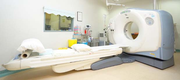
MRI Machine
At the Advanced Neuroscience Summit, I had the incredible opportunity to visit Georgetown's Neuroimaging Center to see a magnetic resonance imaging (MRI) machine in use. Viewing this procedure provided me with a unique experience to view detailed images of structures and tissues of the brain.
This activity also illustrated the ways an MRI can be used and how it functions. An expert in the field walked me through the process of setting up, reading, and working an MRI machine.
2
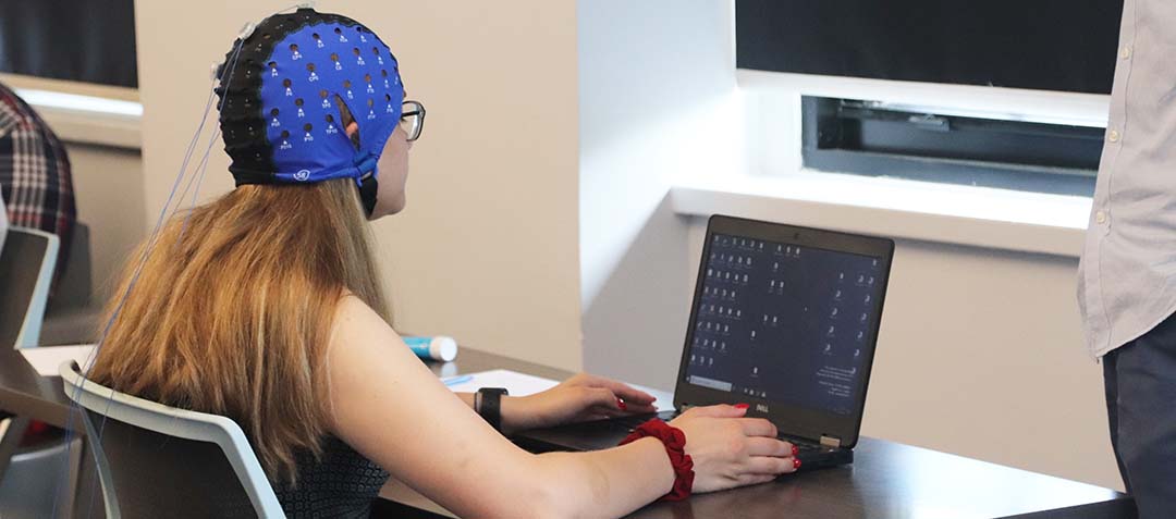
EEG Experience
While at the Summit, I also had the opportunity to see an electroencephalogram (EEG) performed live. This device detects the numerous brain waves that continually occur within the brain. This demonstration showcased the complexity of the brain, and it provided me with the opportunity to view alpha, beta, and theta wave patterns in real-time.
This procedure was performed by a specialist, and it enabled me to ask questions and get real-time feedback.


Manipulating Neurons


Neuropriming
Neural Interfaces

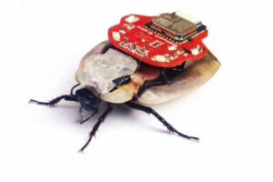
Controlling Cockroaches Through Neural Interfaces
Cockroaches have two antennae on their anterior end. These tiny “hair-like” sensors are connected directly to neurons that communicate messages to the cockroach brain.
Cockroaches have neurons similar to ours, but they have far fewer of them! Cockroaches have only about 1 million neurons, while we have 100 billion.
Even though our nervous systems are very different from a cockroach’s, the structure and function of our individual neurons are quite similar. This allows us to learn about our brains by studying that of a cockroach.
Their neurons communicate just like ours by sending information down their axons in the form of electrical activity called “action potentials”, or as they are referred to in the field, “spikes.”
The more neurons fire, together, the faster pathways are built in your brain. By implanting the tips of tiny electrodes into the cockroach’s antennae, a neural interface is created. Since the electrode wire is electrically conductive, I was able to send artificial electrical signals similar to the one that the cockroach antennae’s sensory neurons naturally create. With this neural interface, the cockroaches could race and move just as I told them because of their electrodes.
Neuroscience Experimentation


Neuropriming Tests Using HALO Devices
Whether you're perfecting your free throw or picking up a new language, you need to form new pathways in your brain in order to learn anything.
The scientific term for this process is called neuroplasticity: your brain’s ability to create and strengthen connections between neurons.
Using specialized equipment, you can create and strengthen motor pathways faster. We call this hyperplasticity or hyperlearning.
The headset works by applying a small electric current to the part of the brain that controls movement, activating neurons so they fire more often when you train.
The more neurons fire, together, the faster pathways are built in your brain. That means you can learn any movement faster — from playing the piano to performing a muscle-up.
When combined with training these devices boost max muscle contraction, fine motor skill acquisition, endurance, and more


Moving Muscles Through Electrical Stimulation
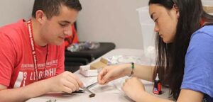
By placing two surface EMG electrodes close to each other across the ulnar nerve, we can manipulate neurons by creating a neural-interface between two people, or even a human and a cockroach!
If people have experienced damage to their peripheral nervous system or spinal cord, their muscles themselves can be stimulated, and this line of research is called "functional electrical stimulation."
For example, functional electrical stimulation can often be used to help someone stand up or to improve walking by helping to swing a foot forward.
This is accomplished by using a spiker box that "copies" the neural impulses of one partner (the controller) and transmits them to the second partner (the control subject).
The more neurons fire, together, the faster pathways are built in your brain. Controlling partner two's movements is possible because the ulnar nerve lies just below the surface of the skin in your forearm and elbow, and it is relatively easy to stimulate.
As a result, the electrical activity from one source is able to catalyze a differing muscle’s contraction, because an external technology brings the two systems together.


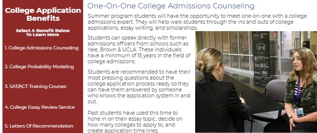

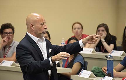
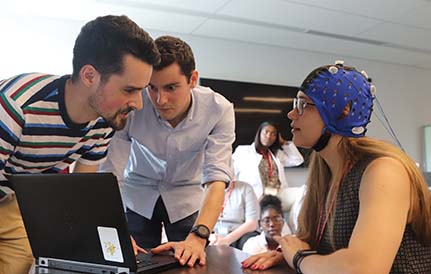
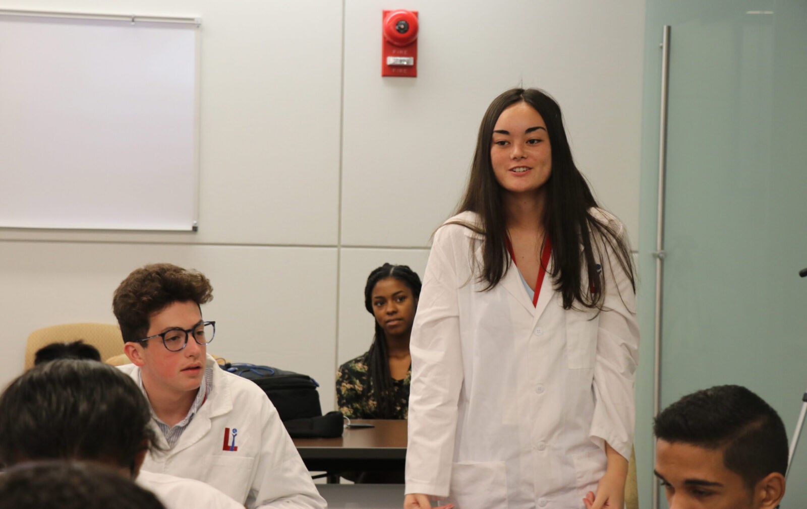
At the Advanced Medical Neuroscience Summit, I had the opportunity to learn at a medical student-level from some of the premier neuroscience professors and doctors in the United States.
When I wasn’t developing my own cutting edge research proposal with the guidance of Dr. Giordano and Dr. Wurzman, I was inspired by the fascinating research presented in lectures structured just as if I were a fellow scientist with experience in research.
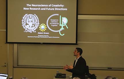
Whether Dr. Green was teaching me about stimulating creativity in the brain, Dr. Ullman was showing the differences in the way kids learn a language, or Dr. Shook was explaining the ethical implications of neuroscience’s future, I got to learn about the most recent neuroscience being discovered. With every graph and conclusion, I realized just how many fields and interests the brain can be applied to.
These panels and lectures made me even more curious about my future in research, and about the complexities of the brain!
Neuroscience Lectures


Deep-Brain Stimulation Viewing
Neuroscience Lectures
Neurological Disorders Panel
Neuroscience Research Proposal
Hospital & Lab Tours
Medical Program Highlights
Click On The Links Below
To See Each Highlight
Neuromodulation, and more specifically, deep brain stimulation, is an increasingly popular surgical procedure most often performed to help people’s symptoms from movement and neurodegenerative disorders. I actually got to watch a real video of the entire procedure. Dr. Giordano explained every step, recounted personal experiences, and paused to point out the most important details.
It felt like I was really in the surgery, but better because I had the chance to really learn why each step was done.
It was so interesting to see the precision of the tools that are used, from drills to electrodes, and how much control the surgeon needed to have. This was one of the activities that really revealed how closely the basic and medical sides of neuroscience are related, going from bench to bedside!
My favorite part was seeing a clip of a patient who stayed conscious while they placed the electrodes in order to make sure they were in the right locations.
Deep-Brain Stimulation Viewing
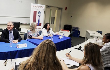
After hearing from people responsible for life-changing science, it was extremely powerful to hear from patients who have had their lives changed for the better. I asked patients and their doctors about their experiences before and after treating their neurological disorders and was able to understand a new perspective. The most incredible part of the panel, was when one of the patients actually turned off his deep brain stimulation device to show all of us the effect it has on his everyday life.
I was so shocked to watch his body language, and his ability to move and talk change so drastically after turning off the device. It was such a noticeable difference, and I felt like it was really clear what these doctors and procedures are capable of.
This was a panel I will remember forever, because I felt such empathy for the patients, and such a sense of power to make positive change.
Neurological Disorders Panel
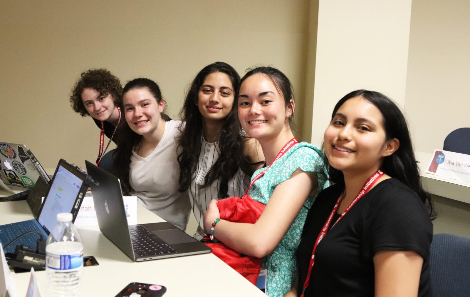
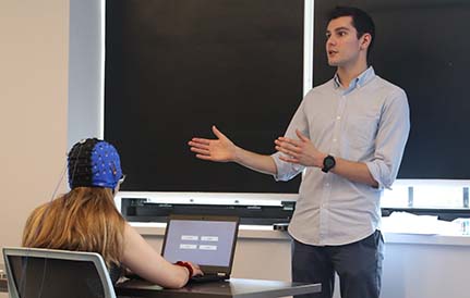
After learning so much about the brain, I worked with a team of students to design a research proposal to address unanswered ethical questions about the future of neuroscience. I used the information from previous lectures as a starting foundation and then conducted research from online publications.
Ultimately, my group landed upon a question that has not been researched in-depth, making our research proposal real cutting-edge neuroethics.
We included in our research proposal a hypothesis, variables, a proposed procedure, what tests we would use, and even investigating the ethical, legal, and social implications of doing our research.
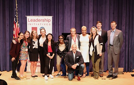
By synthesizing this information into a research proposal, I used the Scientific Method to create a novel idea that could actually be investigated in the future. My team and I presented our proposal to a panel of esteemed doctors, ethicists, and professionals in the field.
Neuroscience Research Proposal
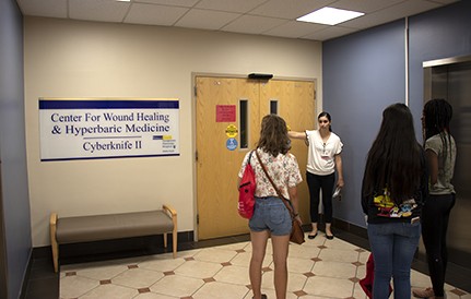
As a result of staying at Georgetown University, I was able to see and experience incredible labs with some of the best technology used in the world. I went on a tour of Georgetown Hospital, where I learned about day-to-day practices and saw the neurological ward and the neurosurgical department. I also went to the Center for Functional and Molecular Imaging at the hospital, where I watched an fMRI being performed and saw their EEG collections. At the Laboratory for Relational Cognition, I watched transcranial alternating current stimulation (tACS) test performed.
At the hospital, I witnessed how the technology operates as the researchers explained what tests they were performing and what role this had in treatment! Finally, at the Petitto Brain and Language Laboratory for Neuroimaging, I got to see how functional infrared spectroscopy works to analyze how young children develop language skills. It’s so impressive how much can be seen in the brain non-invasively, and this helped me develop my research proposal too.
Hospital & Lab Tours

Center For Wound Healing
Georgetown University Medical Center

Our office in Washington, DC is open seven days a week - give us a call! A phone conversation allows us to answer your specific questions and provide you with contact information for previous intership participants and their families so that you can get a first-hand perspective.
Enroll Now
Set Up A Call Today
Join us this summer!

Contact Us
4410 Massachusetts Ave., NW #236
Washington, DC 20016
info@lichange.org
202-738-1115
Helpful LInks
Join A Webinar
Request Alumni References
Become An Intern
Testimonials
Visit The International Internship Homepage
Leadership Initiatives is a 501(c)(3) tax-exempt, publicly supported, charitable organization as determined by the U.S. Internal Revenue Code.


Recognition